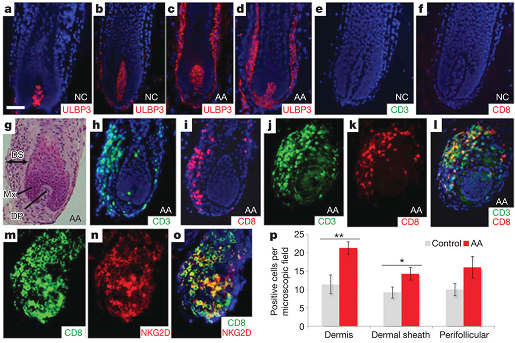
Figure 3. ULBP3 expression and immune cell infiltration of AA hair follicles.
a, b, Low levels of expression of ULBP3 in the dermal papilla of hair follicles from two unrelated, unaffected individuals. c, d, Massive upregulation of ULBP3 expression in the dermal sheath of hair follicles from two unrelated patients with AA in the early stages of disease. e, f, Absence of immune infiltration in two control hair follicles. g, Haematoxylin and eosin staining of AA hair follicle. DS, dermal sheath; Mx, matrix; DP, dermal papilla. h, i, Immunofluorescence analysis using CD3 (h) and CD8 (i) cell-surface markers for T-cell lineages. There is a marked inflammatory infiltrate in the dermal sheath of two affected AA hair follicles. j–l, Double-immunofluorescence analysis with anti-CD3 (j) and anti-CD8 (k) antibodies. l, The merged image clearly shows infiltration of CD3+CD8+ T cells in the dermal sheath of the AA hair follicle. Panels d and g–l are serial sections of the same hair follicle of an affected individual. Counterstaining with 4′,6-diamidino-2-phenylindole is shown in blue (a–f, h, i, l). Scale bar, 50 µm. AA, alopecia areata patients; NC, normal control individuals. m–o, Double immunostainings with anti-CD8 (m) and anti-NKG2D (n) antibodies revealed that most NKG2D+ cells co-expressed CD8+; o, merged image. p, Quantification of immunohistochemical staining (positive cells per microscope field at a magnification of × 200) for ULBP3 in 16 patients with AA and in 7 controls showed a significantly increased number of ULBP3+ cells in the dermis and dermal sheath in patients with AA compared with control skin. In addition, positive cells were also upregulated in perifollicular regions in AA samples, although this was not statistically significant. Data were analysed by Mann–Whitney test for unpaired samples and are expressed as means ± s.e.m.; asterisk, P < 0.05; two asterisks, P < 0.01.

