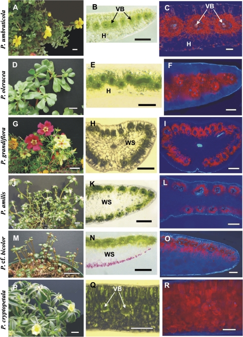
Fig. 1.
General view of Portulaca species (left panels), hand-made leaf cross-sections (middle panels), and distribution of chlorenchyma under the fluorescent microscope (right panels). (A–C) Portulaca umbraticola; (B) distribution of vascular bundles (VBs) in the median paradermal plane; (C) red fluorescence from chlorenchyma is distributed around each VB. (D–F) Portulaca oleracea; (E) zig-zag pattern in the distribution of VBs with their positioning in two paradermal levels, (F) fluorescent chlorenchyma surrounds the VBs. (G–I) Portulaca grandiflora and (J–L) P. amilis; (H, K) VBs are distributed around the leaf periphery, (I, L) chlorenchymatous cells are mostly on the outer side of the VBs. (M–O) Portulaca cf. bicolor; (N) VBs are only on the adaxial side, (O) chlorophyll fluorescence occurs on the adaxial and abaxial sides of VBs. (P–R) Portulaca cryptopetala; (Q) C3-like dorsoventral type of anatomy, (R) chlorenchyma is distributed evenly in leaf section. H, hypoderm; VB, vascular bundles; WS, water storage tissue. Scale bars: 2 cm for A, D, G, J, M, P; 500 μm for B, E, H, K, N, Q; 250 μm for C, F, I, L, O, R.

