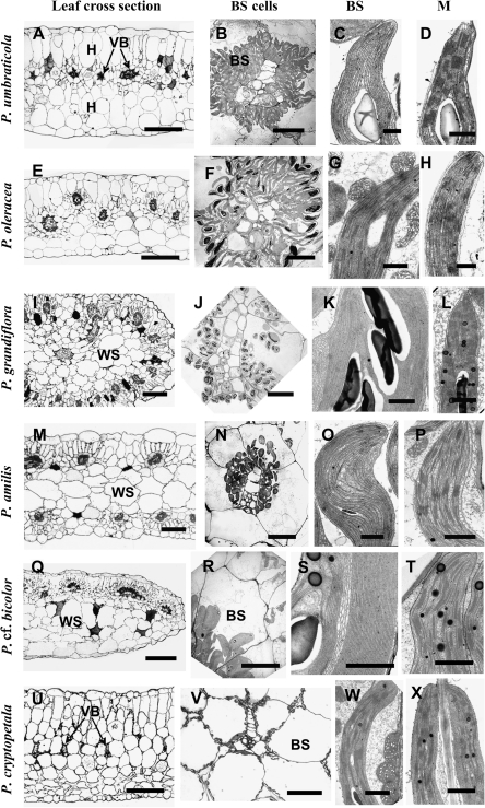
Fig. 2.
Light microscopy of leaf cross-sections, electron microscopy of bundle sheath (BS) cells and of chloroplasts and mitochondria in chlorenchyma cells in Portulaca species. (A–D) Portulaca umbraticola. (E–H) Portulaca oleracea. (I–L) Portulaca grandiflora. (M–P) Portulaca amilis. (Q–T) Portulaca cf. bicolor. (U–X) Portulaca cryptopetala. A, E, I, M, Q, U left panels: light microscopy. B, F, J, N, R, V: BS cells with centripetal positioning of organelles surrounding VBs. BS chloroplasts: (C, K, O, S) grana-deficient (G, W) with well-developed grana and numerous mitochondria. M chloroplasts: (D, L, P, T, X) with well-developed grana and (H) deficient in grana. BS, bundle sheath; H, hypoderm; M, mesophyll; VB, vascular bundle; WS, water storage tissue. Scale bars: 250 μm for A, E, I, M, Q, U; 20 μm for B, F, J, N, R; 100 μm for V; 1 μm for C, D, G, H, K, L, O, P, S, T, W, X.

