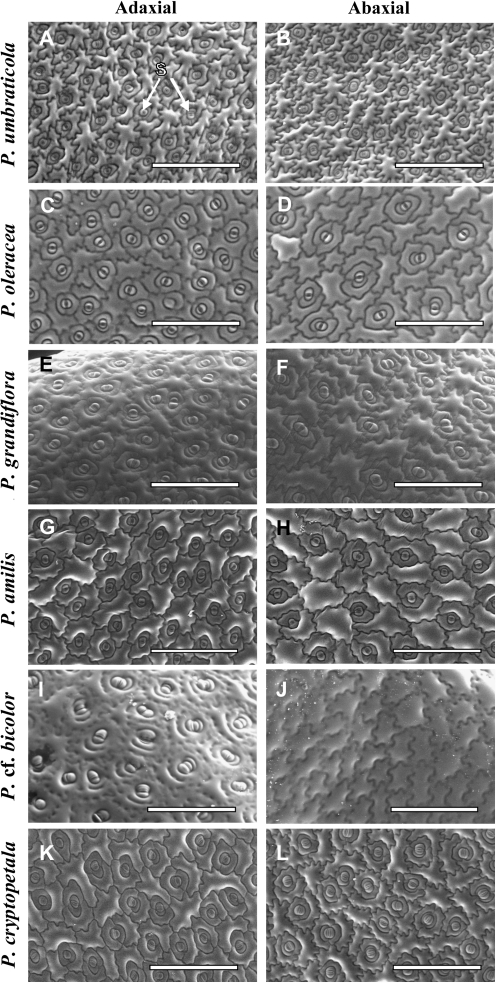
Fig. 6.
Scanning electron micrographs showing stomata distribution on the adaxial (A, C, E, G, I, K) and abaxial (B, D, F, H, J, L) leaf surfaces in six Portulaca species: P. umbraticola (A, B), P. oleracea (C, D), P. grandiflora (E, F), P. amilis (G, H), P. cf. bicolor (I, J), and P. cryptopetala (K, L). S, stomata. Scale bars: 400 μm.

