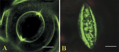
Fig. 4.
Confocal images of stomata labelled by FM1-43. Guard cell pairs of stomata were labelled by FM1-43 and the imaging was performed as described in the Materials and methods. (A) Opened stoma. Note that the plasmalemma is quite smooth and taught. (B) Closed stoma. Note that the ventral plasmalemma becomes folded with numerous extrusions, and excreted vesicles appear nearby, whereas the dorsal plasmalemma becomes invisible.

