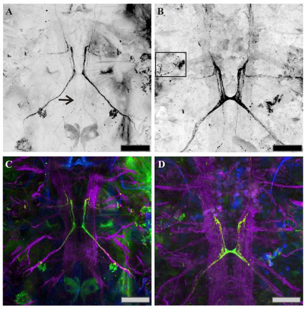
Figure 3.
C-type allatostatin (C-AST)-like and acetylated-α-tubulin-like immunoreactivity complemented by DAPI staining, in developing copepodids. (A, C) Anterior end of a C3 copepodid. Axons from the MBGC-AST are present at the C3 stage (arrow). Somata were not obvious in this preparation in any of the lateral ganglia, but were present in other animals (data not shown). (B, D) By C4, the maxillar commissure is well-formed with projections leading both anterior and posterior from this neuromere. The C-AST-like labeled axons have become larger and longer. (C, D) Along with C-AST antibody (green), these preparations were also labeled with the acetylated-α-tubulin antibody (purple) and a DAPI counterstain (blue) to reveal the nervous system and associated cell nuclei. Scale bars: 50 μm. Images are at brightest pixel projections of 15 optical sections taken at 0.67 μm intervals.

