Figure 1.
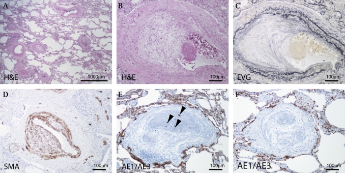
(A) Stenosis and obstruction of many pulmonary arteries and arterioles are shown. Bar, 1000 μm. (B, C) A pulmonary artery showing fibrous thickening of intima and fibrin thrombus (B: H&E stain; C: elastic van Gieson stain). (D) Intimal proliferating fibromuscular cells are positive for α-smooth muscle actin. (E, F) Fibrin thrombus with (E) or without (F) adenocarcinoma cells. Immunoreactivity of pancytokeratin antibody (AE1/AE3) is indicated in the carcinoma cells (F). (E) Arrowheads indicate cancer cells in the vessel. (B–F) Bar, 100 μm.

