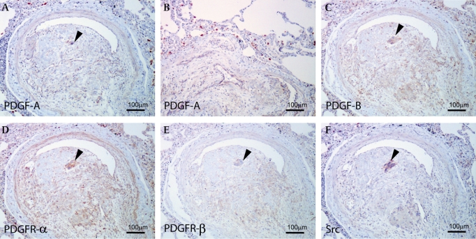
Figure 2.
(A) Carcinoma cells and endothelial cells are immunopositive for platelet-derived growth factor (PDGF)-A. (B) Alveolar macrophages show the overexpression of PDGF-A in the PTTM lesion. (C) Carcinoma cells, endothelial cells and fibromuscular cells are immunopositive for PDGF-B. (D, E) The expression of PGDF receptors (PDGFRs) (PDGFR-α (D); PDGFR-β (E)). PDGFR-α and -β were detected in tumour cells and fibromuscular cells. PDGFR-α were detected in endothelial cells. (F) Immunoreactivity of phosphorylated Src was found in the tumour cells. (A, C, D, E, F) Arrowheads indicate cancer cells in the vessel. (A–F) Bar, 100 μm.

