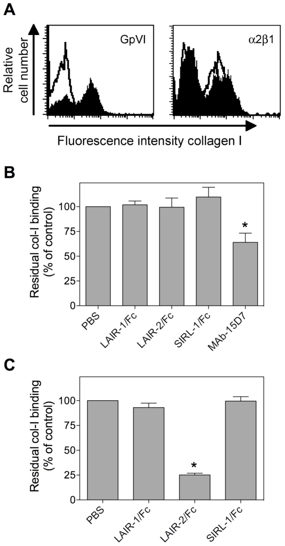
Figure 4. LAIR-2/Fc interferes with GpVI but not α2β1 binding to collagen.
Jurkat-cells expressing GpVI or CHO-cells expressing α2β1 (Mn2+ activated) were incubated with FITC-labeled collagen I (7.7 µg/ml) in the absence or presence of 0.5 mg/ml LAIR-1/Fc, LAIR-2/Fc or SIRL-1/Fc. Residual collagen binding was subsequently analyzed via FACS-analysis. Panel A: representative histograms for binding of FITC-labeled collagen I to GpVI- or α2β1-expressing cells in the absence (closed histograms) or presence (open histograms) of LAIR-2/Fc. Panel B: Quantitative analysis of residual FITC-labeled collagen I binding to α2β1-expressing cells in the absence or presence of Fc-fusion proteins or the blocking α2β1 antibody MAb-15D7. Panel C: Quantitative analysis of residual FITC-labeled collagen I binding to GpVI-expressing cells in the absence or presence of Fc-fusion proteins. Data represent the relative mean fluorescent intensity (expressed as % of PBS control) ± SD of three independent experiments. *p<0.005 compared to control.

