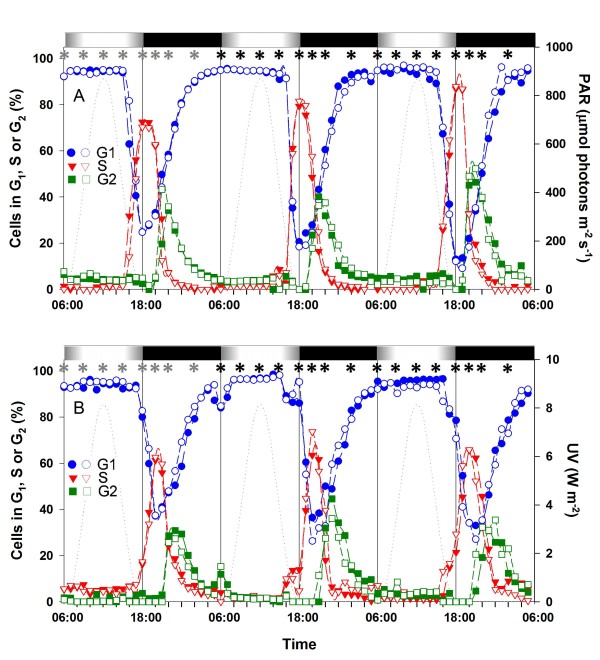
Figure 3.
Effect of UV exposure on the timing of the cell cycle phases of Prochlorococcus marinus PCC9511 cells grown in large volume, continuous cultures used for real time quantitative PCR (qPCR) and microarray analyses. A, distribution of G1 (blue), S (red) and G2 (green) phases for large volume continuous cultures of PCC9511 grown acclimated to HL. B, same for HL+UV conditions. The experiment was done in duplicates shown by filled and empty symbols. Note that only the UV radiation curve is shown in graph B since the visible light (PAR) curve is the same as in graph A. Asterisks indicate the time points of sampling for qPCR (grey) and microarrays (black). White and black bars indicate light and dark periods. The dashed line indicates the growth irradiance (right axis). Abbreviations as in Fig. 1.

