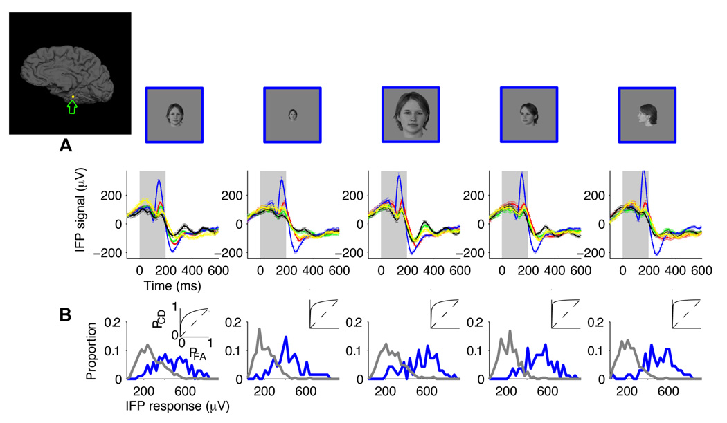
Figure 4. Robustness to scale and depth rotation.
A. Responses of an electrode located in the parahippocampal part of the right medio-temporal-gyrus (Talairach coordinates: 32.6, −34.8, −13.6; see inset depicting the electrode position). Each column depicts the IFP responses to images where the objects were shown at different scales or depth rotations. Each object was presented in a default viewpoint (front view in the case of human faces, see Figure S1B for the “default” viewpoint for the other categories) at a scale of 3 degrees (column 1, total n=329), with the same default viewpoint at a scale of 1.5 degrees (column 2, total n=359), with the same default viewpoint at a scale of 6 degrees (column 3, total n=363), at a scale of 3 degrees and ~45 degree depth rotation (column 4, total n=370) and at a scale of 3 degrees and ~90 degree depth rotation (column 5, total n=355). Above the physiological responses we show an example object to illustrate the changes in scale and viewpoint but the data corresponds to the average over all 5 exemplars for each category. The format for the response subplots is the same as in Figure 1A. Error bars are SEM. The gray rectangle denotes the stimulus presentation time. Figure S8 shows the responses to each one of the exemplars for this electrode. B. Distribution of IFP responses (IFP range in the 50 to 300 ms window) for “human faces” (blue) versus other object categories (gray). The insets show the corresponding ROC curves.

