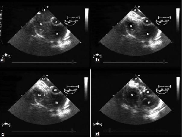
Figure 1.

Transesophageal echo in the short axis view of the aorta (a) showing a large secundum atrial septal defect (b) Successful closure of the defect using 17 mm Amplatzer septal occluder (c) Migration of the device 24 hours later at the aortic end in the anteroposterior plane. The migrated device was successfully retrieved percutaneously, (d) Successful deployment of the 20 mm Amplatzer septal occluder
