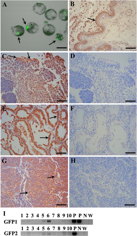
Figure 1.
Contribution of porcine SKPs into neural and mesodermal lineages. (A) eGFP expression in injected day 5 IVF embryos 24 h post injection. (B) Positive control for anti-eGFP antibody: the section was from a uterus where placenta tissue was GFP positive (arrow) while epithelial endometrium was eGFP negative. This control fetus was created by nuclear transfer with eGFP transgenic donor cells. (C-H) Immunofluorescent staining of brain (C), kidney (E) and genital ridge (G) of chimeric fetuses. The corresponding negative controls with only secondary antibody were shown: brain (D), kidney (F) and genital ridge (H). The arrows show representative eGFP positive cell areas. (I) PCR results using genomic DNA from various tissues: brain (1-2), skin (3-4), kidney + genital ridge (5-6), liver (7-8) and body trunk (9-10). P: positive controls using the plasmid pEGFP-N1 as the template; N: negative control without template. W: wild-type porcine genomic DNA. GFP1 and GFP2 are different primer sets (Table 2). One fetus is in lanes 1, 3, 5, 7 & 9; while the other fetus is in lanes 2, 4, 6, 8 & 10. Scale bars: 100 μm (A); 50 μm (B-H).

