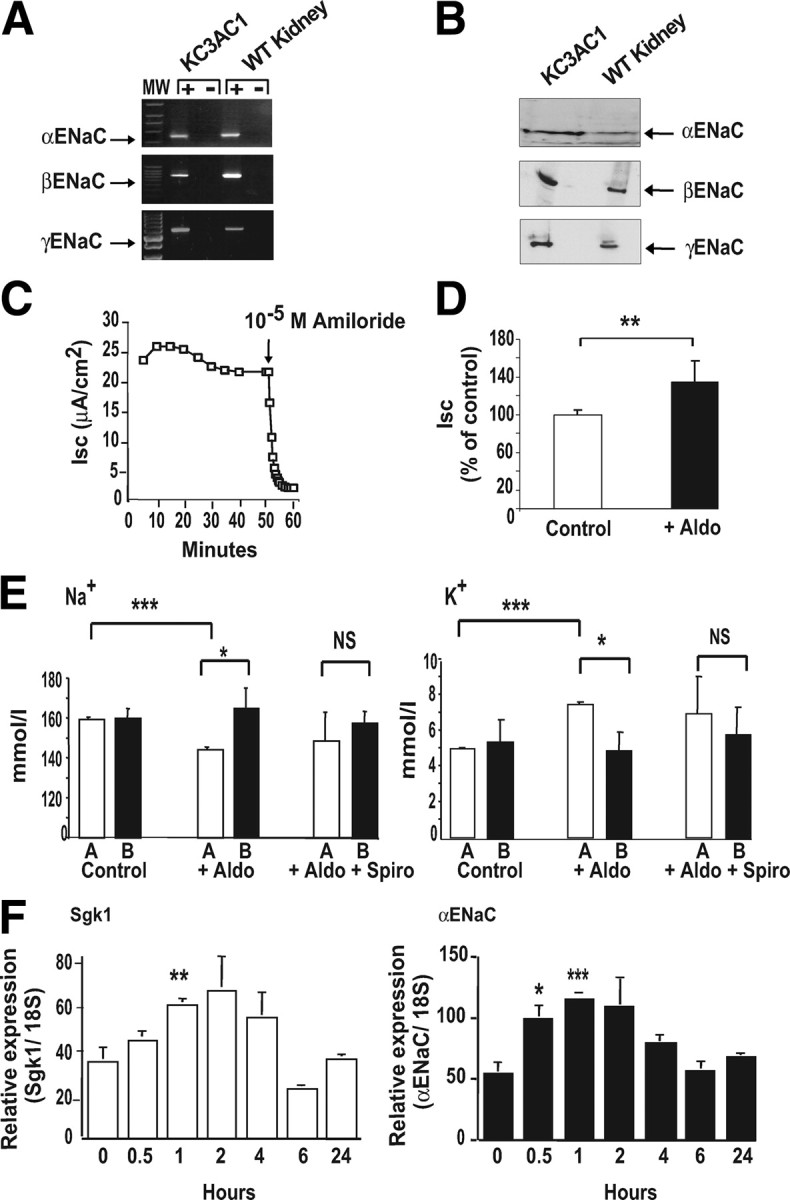
Fig. 2.

Aldosterone stimulates Na+ reabsorption in KC3AC1 cells through ENaC. A, RT-PCR was performed as described in Materials and Methods and allowed the detection of a 555-bp (aENaC), a 631-bp (βENaC), and a 670-bp (gENaC) amplicon. B, Western blot analysis allowed the detection of the three subunits of ENaC at 90 kDa (αENaC), 92 kDa (βENaC), and 85 kDa (γENaC). C, electrophysiological studies. Cells were grown on Snapwell filters in MM for 24 h. The short-circuit current (Isc, μA/cm2), transepithelial voltage (VT, mV) and transepithelial resistance (RT, Ω/cm2) were measured as described previously (29 ). When 10−5 m amiloride was applied at the apical side of the KC3AC1 cells, the short-circuit current was almost completely abolished within minutes. D, Cells, cultured on Snapwell filters as described above, were incubated for 18 h in the absence (Control) or presence of 10−8 m aldosterone (+Aldo). Each point represents mean ± sem of at least three independent determinations performed in duplicate or in triplicate. Statistical significance: **, P < 0.01. E, Ionic measurements. Cells were seeded on collagen I-coated Transwell filters and were cultured for 5 d in the epithelial medium. Cells were rinsed with PBS then were cultivated in MM for 24 h in the presence of 10−8 m aldosterone (+Aldo) or 10−8 m aldosterone + 10−6 m spironolactone (+Aldo + Spiro). A = apical; B = basolateral. Four hundred microliters of the supernatant was recovered from the medium bathing the apical and basolateral surface of the KC3AC1 cells. Na+ (left) and K+ ions (right) were then measured by the “Centre d’Experimentation Fontionnelle Intégrée” of the IFR Claude Bernard (Paris, France) on an Olympus automate. Data represent means ± sem of three independent determinations. Statistical significance: *, P < 0.05, and ***, P < 0.001. F, qRT-PCR analysis of sgk1, and αENaC mRNA expression in KC3AC1 cells after aldosterone treatment. Highly differentiated KC3AC1 cells were grown on filters for 24 h in MM and subsequently treated for various periods of time (0, 0.5, 1, 2, 4, 6, or 24 h) with 10 nm aldosterone. Results were normalized by the amplification of 18S RNA and are expressed as relative fold induction compared with basal conditions. Data represent means ± sem of at least three independent determinations performed in duplicate. Statistical significance: *, P < 0.05; **, P < 0.01; and ***, P < 0.001.
