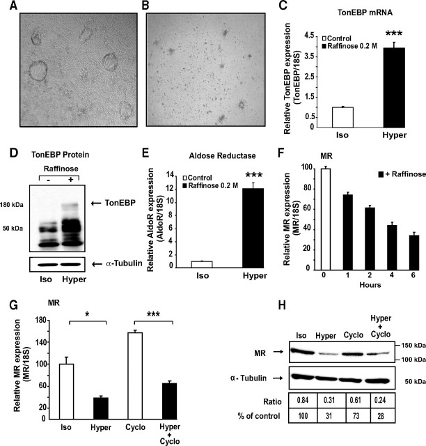
Fig. 4.
Hypertonic stress inhibits MR expression. A, B, Phase-contrast micrographs of a monolayer of differentiated KC3AC1 cells incubated in the absence (A) or in presence of 0.2 m raffinose (B) for 6 h. Note the absence of domes in raffinose-treated cells. C, E, Hypertonicity stimulates TonEBP and aldose reductase expression. Differentiated KC3AC1 cells were incubated in the absence or the presence of 0.2 m raffinose for 6 h. mRNA expression was measured by qRT-PCR. Results are expressed as attomoles/femtomoles of 18S are mean ± sem of 30–36 independent determinations. Statistical significance: *, P < 0.05; **, P < 0.01; ***, P < 0.001. D, Western blot analysis of TonEBP in KC3AC1 cell lysates exposed for 18 h to raffinose. Fifteen micrograms of protein from KC3AC1 cell homogenates were processed for immunoblotting with anti-TonEBP SHN50 antibody. Note the presence of a specific band of approximately 180 kDa in KC3AC1 cell lysates exposed to raffinose. F, G, Analysis of MR transcript by qRT-PCR. F, Differentiated KC3AC1 cells were incubated for various periods of time in the absence or presence of 0.2 m raffinose. G, Differentiated KC3AC1 cells were incubated for 6 h in the absence (Iso) or in the presence of 0.2 m raffinose (Hyper) and/or cycloheximide (Cyclo, 5 μg/ml). Total RNA was extracted and processed for qRT-PCR as described in Fig. 3. Data represent means ± sem of at least three independent determinations performed in duplicate. Statistical significance: *, P < 0.05 and ***, P < 0.001. H, Western blot analysis of MR expression in KC3AC1 cells. Differentiated KC3AC1 cells were incubated for 18 h in the absence (Iso) or in the presence of 0.2 m raffinose (Hyper) and/or cycloheximide (5 μg/ml). Western blot analyses were performed as described in Fig. 3.

