Fig. 6.
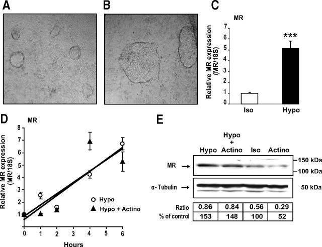
Hypotonic stress stimulates MR expression. A and B, Phase-contrast micrographs of monolayers of differentiated KC3AC1 cells incubated for 6 h with isotonic (A) or a hypotonic medium (B). Note that the domes seemed larger in KC3AC1 cells incubated under hypotonic conditions. C and D, Analysis of MR transcripts by qRT-PCR. C, Differentiated KC3AC1 cells were incubated for 6 h with isotonic (Iso) or a hypotonic medium (Hypo). D, Differentiated KC3AC1 cells were incubated for various periods of time under hypotonic conditions (Hypo) and/or in the presence of 0.4 μm actinomycin D (Actino). Total RNA was extracted and processed for qRT-PCR as described in Fig. 3. Statistical significance: ***, P < 0.001. E, Western blot analysis of MR expression in KC3AC1 cells. Differentiated KC3AC1 cells were incubated for 18 h with isotonic (Iso) or hypotonic medium (Hypo) and/or in the presence of 0.4 μm actinomycin D (Actino). Western blot analyses were performed as described in Fig. 3.

