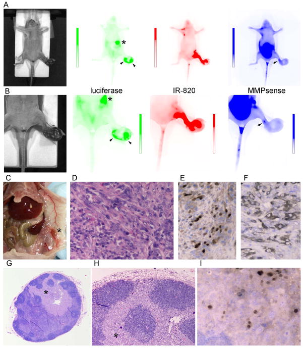
Figure 5. Multiplex Optical Imaging of a Hairless Rhabdomyosarcoma Model.
A, From left to right: photograph, luciferase luminescence (scale 2.8×10⁵ to 8.0×10⁸ p/sec/cm2/sr), IR-820 fluorescence (scale 3.2×10⁵ to 1.6×10⁹ p/sec/cm2/sr) and MMPsense fluorescence (scale 0 to 2.8×10⁹ p/sec/cm2/sr) for a mouse bearing a rhabdomyosarcoma of the left foot. Viable tumor is shown with arrowheads. A metastatic lesion is shown by an asterisk (*). MMPsense activity is shown by an arrow at the advancing front of the tumor. Apparent MMPsense signal in the abdomen is artifact attributable to alfalfa in mouse chow. B, repeat imaging for a more restricted field of view. Scales: luminescence (2.8×10⁴ to 8.6×10⁸ p/sec/cm2/sr), IR-820 fluorescence (2.1×10⁷ to 3.4×10⁹ p/sec/cm2/sr) and MMPsense fluorescence (0 to 4.9×10⁹ p/sec/cm2/sr). C, necropsy photomicrograph demonstrating an enlarged subcutaneous lymph node that overlied the spleen. D, H&E of primary tumor consistent with rhabdomyosarcoma (400×). E to F, Immunohistochemistry positive for nuclear myogenin and cytoplasmic desmin, respectively (400×). G, H&E of lymph node shown in A, B and C (20×). H, Close-up of G (200×). I, Myogenin positive rhabdomyosarcoma cells in the lymph node (400×).

