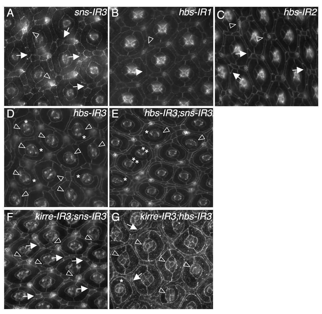
Figure 3.

Sns and Hbs act redundantly in patterning ommatidia. Eyes at 42 h APF were stained with an antibody against either Armadillo (B–C), E-cadherin (A, D–F) or Echinoid (G). (A) Strong reduction of Sns by sns-RNAi (sns-IR3). Frequently, single cells failed to be selected at vertices (arrows). Occasionally, a cluster of cells was found surrounding a bristle group (arrowheads). (B) Mild reduction of Hbs by expressing a single copy of hbs-RNAi (hbs-IR1). A mis-positioned cell is highlighted by an arrow and a cluster of cells surrounding a bristle group indicated by an arrowhead. (C) Expression of two copies of hbs-RNAi (hbs-IR2). Single cells were not selected in vertices (arrows) and bristle groups misplaced (arrowheads). (D) Strong reduction of Hbs by hbs-RNAi (hbs-IR3). Defects in cone cells (arrowheads) and 1°s (asterisks) are highlighted. (E) Strong reduction of both Hbs and Sns. Ommatidia in direct contact are indicated by double asterisks. Defects in cone cells (arrowheads) and 1°s (asterisks) are highlighted. IOCs completely failed to sort into single file. (F) Strong reduction of both Kirre and Sns. Single cells failed to be selected within vertices (arrows). Extra cells (‘cone contact cells’) were commonly found in direct contact with cone cell quartets (arrowheads). (G) Strong reduction of both Kirre and Hbs. Frequently cone cells formed abnormal configurations (arrowheads). Occasionally IOCs failed to sort into single rows (arrows).
