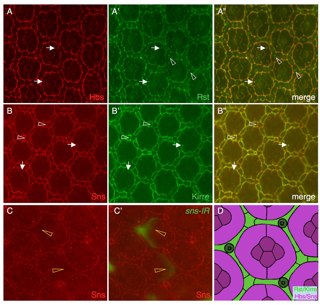
Figure 4.

IRM proteins are expressed in complementary cell types. Eyes at 27 h APF are shown in A–B and an eye at 42 h APF in C. (A-A”) Hbs (red) and Rst (green) co-localize on the surface. Hbs also co-localizes with Rst in vesicles in 1°s (arrows). Rst is also found in vesicles in IOCs (arrowheads), where it does not co-localize with Hbs. A merged view is shown in A”. (B-B”) Sns (red) co-localizes with Kirre (green) on the cell surface. Sns and Kirre vesicles largely co-localize (arrows). Sns and Kirre were also found at the borders between IOCs and bristle groups (arrowheads). (C-C’) sns-RNAi (sns-IR3) was targeted to single cells (green, C’) and the eye was stained with an anti-Sns antibody (red). Sns is reduced at the border when sns-IR is targeted to 1°s (arrowheads). D) Schematic representation of expression domains of the IRM proteins. Hbs and Sns of the Nephrin group (magenta) are expressed 1°s and cone cells. Kirre and Rst of the Neph1 group are expressed in IOCs (green).
