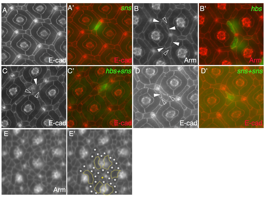
Figure 6.

Sns and Hbs drive separation of cells from ommatidia. Eyes at 42 h APF were stained with an antibody against either Armadillo (Arm) or E-cadherin (E-cad) as indicated. Merged views in A–D are shown in A’-D’. (A-A’) When a single copy of sns (green) was expressed in a single cell, the cell retained its normal position. (B-B’) Upon expression of hbs (green), two cells were separated from one but retained contact with another ommatidium. Wild type IOCs maintained small interfaces with each other (open arrowhead) while target IOCs established larger interfaces with IOCs (arrowheads). (C-C’) When both hbs and sns were expressed in a single cell (green), the cell was fully separated from ommatidia. Wild type IOCs maintained small interfaces with each other (open arrowheads) but larger interfaces with the target IOC (arrowhead). (D-D’) When two copies of a sns transgene were expressed in a single cell (green), the cell was fully separated from ommatidia. Wild type IOCs maintained small interfaces with each other (open arrowheads) but larger interfaces with the target IOC (arrowhead). (E-E’) Pupal eye at 20 h APF. The eye was stained with an anti-Armadillo antibody. Emerging 1°s are outlined (E’). Most IOCs (white asterisks) are found in touch with at least one 1°. In the same area, an IOC (gold asterisk) not in touch with any 1° is highlighted.
