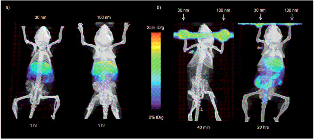
Figure 12.
MicroPET/CT studies at various times after injecting the mice with 30 and 100 nm 64Cu-labeled SNPs. a) In vivo biodistribution studies of the SNPs by systemically injecting the SNPs into mice through the tail veins. Left panel: 30 nm SNPs; right panel: 100 nm SNPs. b) Lymph node trafficking studies of SNPs using front footpad injections. The 30 and 100 nm SNPs were injected on different sides of footpads of a mouse. MicroPET/CT was carried out for 40 min immediately after injection (left) and 20 h after injection (right). From Ref.[100].

