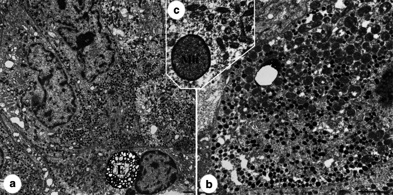
Fig. 5.
Electron microscopy image of silent corticotroph adenoma. a Low power view of case 10 shows polygonal cells with numerous mitochondria, pleomorphic granules, and enigmatic body (E). Higher magnification b highlights increased mitochondrial density and variability in the size and electron density of granules. Case 18 showed mitochondrial enlargement and peripheral condensation of matrix (Mit on c). Bars 2 μm.

