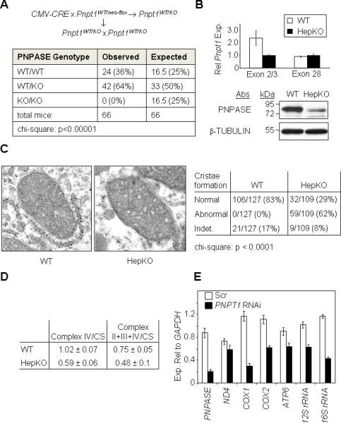
Figure 1. Deletion of Pnpt1 in Hepatocytes Impairs Mitochondrial Function.

(A) Breeding strategy (top) and results (bottom) for attempting to generate a PNPASE KO mouse.
(B) Hepatocyte-specific Pnpt1 KO (HepKO) in 4-week old mice. Top: QPCR for liver Pnpt1 expression using an exon 2 – exon 3 primer pair versus a primer pair within exon 28. Bottom: PNPASE immunoblot from 4-week old WT and HepKO mouse livers.
(C) HepKO mitochondria have altered cristae. Left: TEM of 6-week old littermate livers shows circular, smooth HepKO IM cristae in contrast to linear, stacked cristae of WT mitochondria. Right: Analysis of cristae morphology in which a single normal cristae within a mitochondrion was scored as normal. Indet = indeterminate.
(D) Decreased respiration in isolated HepKO mitochondria. Oxygen consumption (nmol/min/mg protein) for ETC complexes IV and II+III+IV was measured using an O2 electrode. Mitochondrial mass was determined by citrate synthase (CS) activity using a spectrophotometer. Respiratory activities are shown normalized to CS activity.
(E) Decreased mature mtRNAs in HEK293 cells with RNAi to PNPT1. Transcripts were quantified relative to cytosolic GAPDH expression by QPCR from HEK293 cells 7d post-infection (nadir) with scramble (Scr) or PNPT1 RNAi retroviral constructs. See also Figure S1, S2, and S3.
