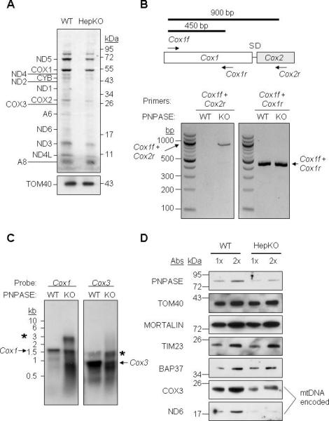
Figure 2. HepKO Liver Mitochondria Do Not Efficiently Process mtRNA Precursors.

(A) In organello protein synthesis. WT and HepKO mitochondria (100 μg) were treated with micrococcal nuclease S7, and in organello translation was performed using [35S]-MET. TOM40 immunoblot shows equivalent mitochondria in each assay.
(B) RNA was isolated from WT and HepKO liver mitochondria followed by DNase I treatment to remove contaminating DNA. RT-PCR was performed for Cox1 and Cox2 with primers shown in the schematic (upper) and separated on a 1.5% agarose gel.
(C) Northern blot of mtRNA from WT and HepKO mouse liver mitochondria using a Cox1 or Cox3 DNA probe. * marks larger precursor mtRNAs and the arrow shows the mature mtRNA.
(D) Steady-state expression of nuclear- and mitochondrial-encoded proteins in WT and HepKO liver mitochondria. Equivalent nuclear-encoded protein expression shows that HepKO reduced mitochondria-encoded protein expression was not due to differing mitochondrial content between WT and HepKO liver cells. See also Figure S4.
