Figure 8.
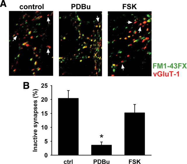
PDBu reduces the percentage of inactive excitatory synapses. A, Example images from control (left), PDBu (middle)-, and FSK (right)-treated synapses. Active synapses were labeled with FM1-43FX (green); glutamatergic terminals were identified by immunoreactivity to vGluT-1 antisera (red). Puncta that are positive for vGluT-1 antisera but devoid of FM1-43FX labeling are inactive excitatory synapses (arrows). B, Quantification of the percentage of inactive glutamatergic terminals in control (20.4 ± 2.8%), or PDBu (3.6 ± 1.1%)-, and FSK (15.2 ± 3%)-treated synapses (*p < 0.00005 compared with control; n = 5). Summary results represent 250 puncta in each condition sampled in 50 fields from five independent experiments. Fields were selected and analyzed by an observer naive to experimental condition. Error bars indicate SEM.

