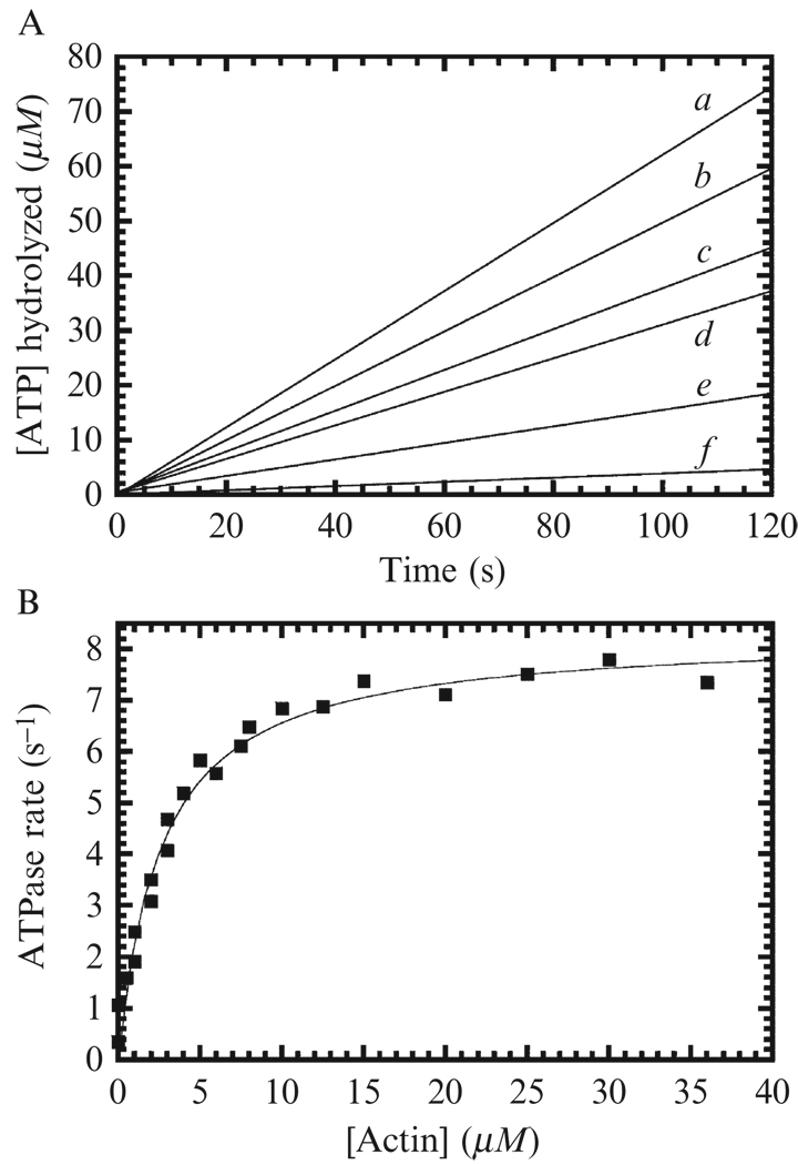
Figure 6.2. Steady-state ATPase activity of myosin VI.
(A) Time course of ATP turnover by 100 nM myosin VI at 16 (a), 8 (b), 4 (c), 2 (d), 1 (e), and 0 µM (f) actin filaments using the NADH-coupled assay. (B) Actin filament concentration dependence of the steady-state turnover rate of myosin VI. The solid line through the data points is the best fit to Eq. (6.1), with kcat = 8.3 ± 0.2 s−1 and KATPase = 2.8 ± 0.3 µM. Data are from (De La Cruz et al., 2001).

