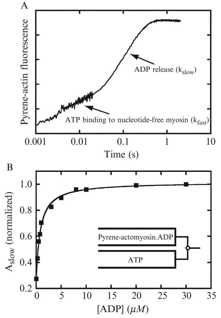
Figure 6.6. ADP dissociation determined by ATP-induced dissociation of pyrene-actomyo1b.
(A) Pyrene fluorescence transient obtained by mixing 0.15 µM myo1b equilibrated with 2 µM ADP with 1 mM ATP. The time course is presented on a log scale to show the slow and fast exponential phases. (B) Normalized amplitude of the slow phase obtained by fitting pyrene transients to double exponential functions (Eq. (6.7)) as a function of ADP concentration. The solid line is a fit of the data to Eq. (6.12). Data are from (Lewis et al., 2006).

