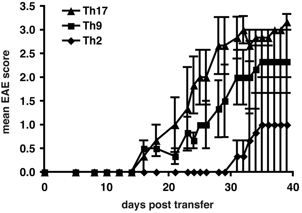
Figure 6. Induction of EAE with secondary Th17, Th9 and Th2 cells.
Secondary Th9 and Th17 cells induce EAE upon adoptive transfer, while secondary Th2 cells induce no or mild/delayed disease. MOG-specific CD4+CD62Lhi T cells were stimulated with irradiated APCs and MOG35-55 and differentiated in vitro into Th17, Th9 or Th2 cells with polarizing cytokines. Th17 cells were supplemented with IL-23. Th9 and Th2 cells were supplemented with IL-2. After 6 days cells were re-stimulated with anti-CD3 and anti-CD28 Abs for 48h without cytokines. 5×10⁶ cells were injected i.v. into C57Bl/6 recipients that received pertussis toxin. Recipient animals were observed for the development of clinical signs of EAE for 40 days. Shown are the mean clinical scores of one experiment, error bars represent SEM. Similar results were obtained in 2 independent experiments.

