Figure 6.

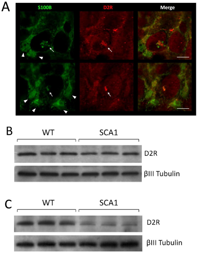
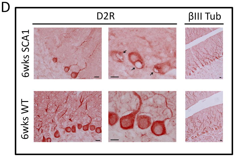
(A) 5 week old SCA1 Tg mice were perfused as previously described (Vig et al. 2009) and 50 μm vibratome sections were immunostained for D2R and S100B. S100B immunostaining is shown in green and D2R in red. Triangles point to S100B positive Bergmann Glia and arrows point to S100B and D2R positive PC vacuoles. Scale bar: 10μm. (B) 4 week old and (C) 6 week old SCA1transgenic mice and WT mice cerebellar extracts were subjected to western blotting loading 25μg per well followed by detection with D2R and βIII Tubulin antibodies. (D) 6 week old SCA1 Tg mice and WT mice cerebellar sections were immunostained with D2R and βIII Tubulin antibodies followed by HRP secondary staining. D2R protein levels are visibility reduced in SCA1 PCs compared to WT, although the number of βIII Tubulin positive PCs remains constant. Arrows point to SCA1 PC vacuoles. Scale bar: 10μm.

