
Figure 1.
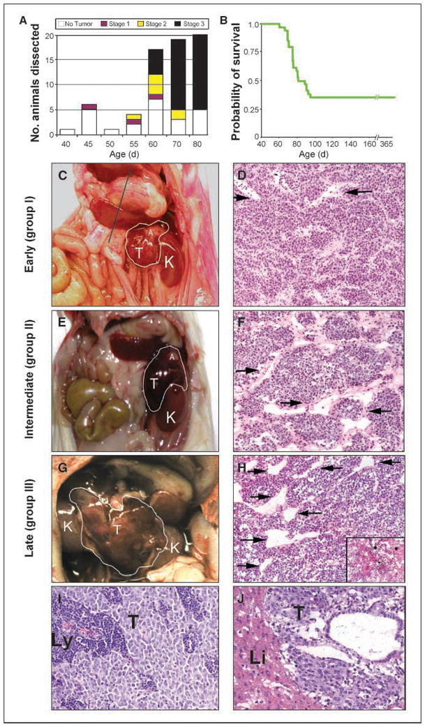
Malignant progression of neuroblastoma in hemizygous mice transgenic for TH-MYCN. A, bar graph of animals dissected at specific time points illustrating the incidence of localized, intermediate, and advanced tumors. B, Kaplan Meier analysis shows that 65% of mice transgenic for TH-MYCN died of tumor by 95 d. C, E, G, gross appearance and grouping of murine tumors at representative ages. D, F, H, H&E staining illustrates similar histopathology for tumors at all groups. Vessel caliber was increased in intermediate and advanced tumors compared with localized tumors (arrows). Regionally spread tumors showed increased necrosis compared with localized tumors (inset in H). I, J, H&E staining showing localized spread to lymph nodes in a group I tumor (I) and distant metastasis to liver in a group III tumor (J). K, kidney; T, tumor; A, adrenal gland; Li, liver; Ly, lymph node.