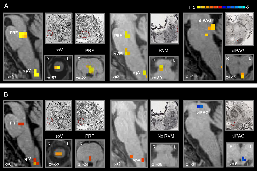
Figure 3. Brainstem differences of brush to either the primary or secondary area versus the normal skin.
Group statistical comparison t maps (radiological convention; p<0.05, corrected), overlaid on high resolution anatomical images in Talairach space, showing in 11 healthy subjects significant differences in the brainstem during brush to either (A) the primary or (B) secondary area following heat/capsaicin application to the right ophthalmic division of the trigeminal nerve versus brush to the untreated skin of the same cutaneous territory. Sketches and micrographs adapted from the atlas of Duvernoy (1995) were used to help identify the location of activation clusters.
(spV = spinal trigeminal nucleus; PRF = pons reticular formation; RVM = rostroventromedial medulla; dlPAG = dorsolateral PAG; vlPAG = ventrolateral PAG).

