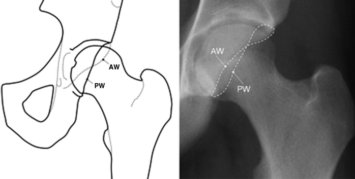
Figure 7.
Focal acetabular over-coverage. Acetabular retroversion is visualized on AP pelvic radiographs by carefully tracing the anterior (AW) and posterior (PW) walls of the acetabular fossa to form the ‘crossover sign.’ A normal acetabulum is anteverted with the anterior rim projecting medial to the posterior rim (see Figure 6). In a retroverted acetabulum, the anterior rim projects lateral to the posterior rim proximally and crosses over in a medial direction distally. (Source: Reprinted with permission M. Tannast, K.A. Siebenrock, S.E. Anderson, Femoroacetabular Impingement: Radiographic Diagnosis – What the Radiologist Should Know, AJR Am J Roentgenol, 188(6), p. 1545, © 2007 American Roentgen Ray Society.)

