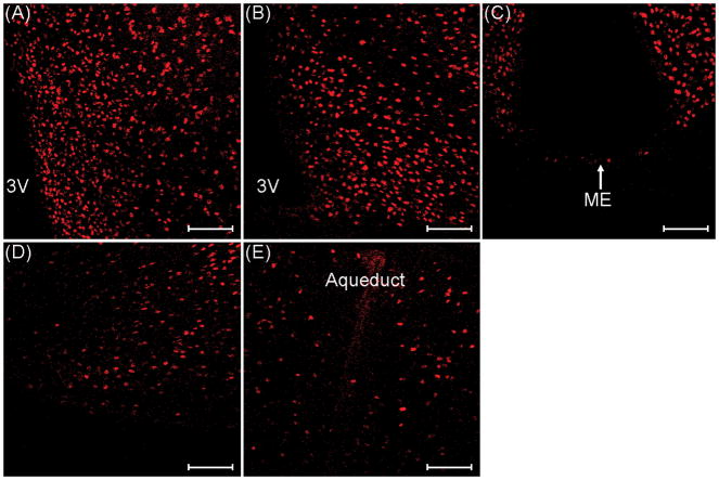
Fig. 2.
TOP-immunoreactive cells in the A) medial preoptic area, B) arcuate nucleus, C) median eminence (ME), D) ventrolateral portion of the ventromedial nucleus of the hypothalamus, and E) midbrain central grey of ovariectomized control mice. 3V, third ventricle. Images were taken at 200× magnification. Scale bars = 100 μm.

