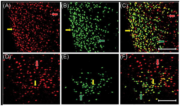
Fig. 4.
Cells in the medial preoptic area (mPOA, panels A–C) and ventrolateral portion of the ventromedial hypothalamic nucleus (VMNvl, panels D–F) coexpress TOP (red) and ERα (green) from a control animal. TOP alone (A and D), ERα alone (B and E) and overlaid image of cells expressing both TOP and ERα (C and F). Cells in the mPOA and VMNvl expressing TOP only (red arrows), ERα only (green arrows) and both TOP and ERα (yellow arrows). Images were taken at 200× magnification. Magnification bars = 100 μm. 3V, third ventricle.

