
Figure 4.
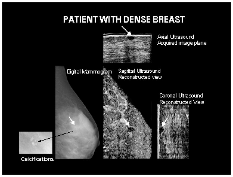
A benign circumscribed oval hypoechoic mass in a 36 year old patient with dense breast tissue, identified by white arrows, is seen on the acquired and reconstructed ultrasound planes. The mass was occult on the mammogram. Shown in the insert corresponding to the mammogram are benign calcifications not visible on the ultrasound images.