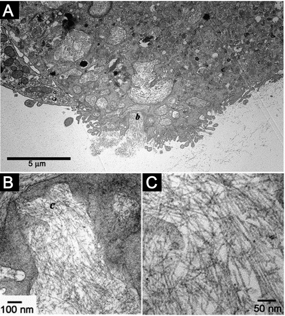
Figure 7.
(A) TEM micrograph of a cell entrapped in the nanofibrillar matrix internalizing the PA nanofibers. (B) Intermediate magnification of the region marked b in (A) showing the formation of intracellular membrane delineated compartments filled with nanofibers. (C) High magnification micrograph of the nanofibers in the area marked c in (B)20.

