
Figure 4. DIDS and WFA bind specifically to the VDR LBD.
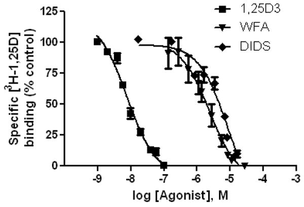
Radiolabeled steroid competition was used to generate VDR IC50 curves for 1,25D3, WFA and DIDS (see methods). The curves represent data pooled from three separate experiments where the concentration of protein was held constant. The IC50 values for 1,25D3, WFA and DIDS were measured to be ~8 nM, 2 μM and 8 μM respectively.