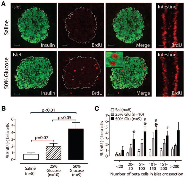
FIG. 3.
Glucose infusion increases β-cell replication in a dose-dependent fashion. A: Representative islets stained for BrdU and insulin show glucose-induced replication; intestine is a positive control for BrdU exposure and staining. Inset: Confocal microscopy confirms BrdU-positive nuclei belong to insulin-positive cells. B: 50% glucose increases β-cell replication (vs. 25% glucose or saline); 25% glucose also shows a strong trend (vs. saline). C: Glucose (Glu) induces more replication in midsize islets than small islets. *P < 0.05 vs. saline; #P < 0.05 vs. “<20” islets. Scale bars = 20 μm; mean ± SE. Sal, saline.

