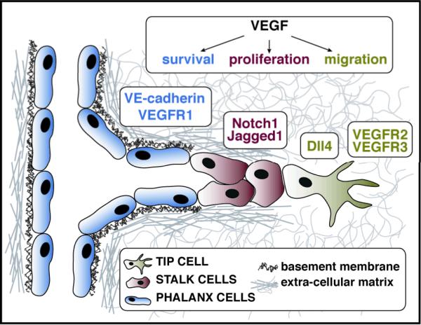
Figure 1.
Endothelial cells in a growing vascular sprout are functionally and molecularly distinct. A growing sprout is composed of tip cells (green), stalk cells (red), and phalanx cells (blue). Each cell type is characterized by a unique molecular signature, resulting in a differential response to VEGF. Tip cells exhibit a migratory response to VEGF, and show an upregulation of Dll4, VEGFR3, and VEGFR2. Stalk cells undergo proliferation and show upregulation of Notch1 and Jagged1. VEGF signaling in phalanx cells leads to a survival response mediated by increased levels of VE-cadherin and VEGFR1.

