
Figure 1.
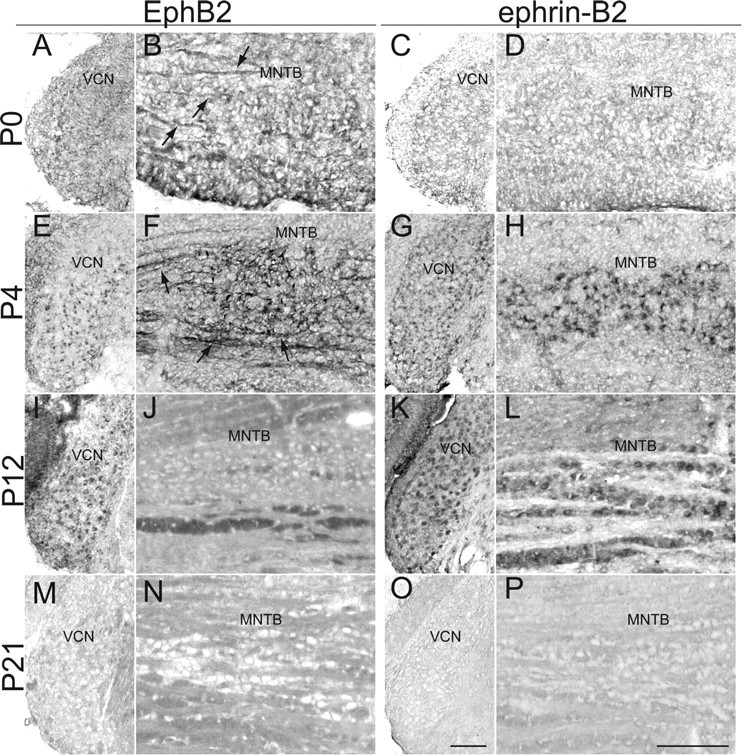
Expression of EphB2 and ephrin-B2 during postnatal development. At P0: A, EphB2 is lightly and evenly expressed throughout VCN. B, EphB2 is also expressed in VCN projection axons (arrows) in the brainstem near MNTB. C, Ephrin-B2 expression is not evident in VCN at P0. D, Ephrin-B2 is not expressed in MNTB. At P4: E, EphB2 expression in VCN is stronger and more punctate than at P0. F, EphB2 expression in VCN axons (arrows) and VCN axon terminals is stronger than at P0. G, Ephrin-B2 shows a more punctate expression pattern than at P0. H, Ephrin-B2 expression appears to be perisomatic in MNTB. At P12: I, EphB2 expression in VCN remains strong and punctate. J, EphB2 is no longer expressed in VCN axons. K, Ephrin-B2 expression remains strong and punctate in VCN. L, Similar to P4, ephrin-B2 staining appears to be perisomatic in MNTB. At P21: M, EphB2 is not expressed in VCN. N, There is a lack of EphB2 staining in VCN axons in the brainstem. O, There is a lack of ephrin-B2 staining in VCN. P, There is no apparent ephrin-B2 staining in MNTB. Scale bars: O (for A, C, E, G, I, K, M, O), P (for B, D, F, H, J, L, N, P), 100 μm.