Figure 4.
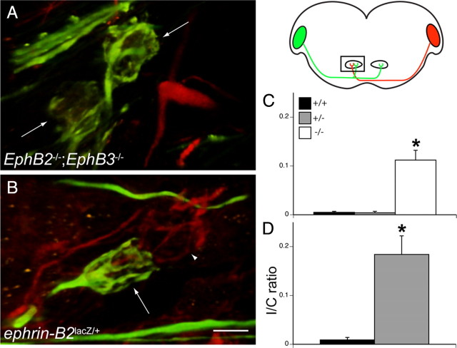
Aberrant projections persist after postnatal day 20. The schematic diagram on the right indicates orientation and dual dye labeling protocol. Alexa Fluor 488 dextran amine (green) was applied to one VCN, and RDA (red) was applied to the other VCN. In the diagram, the rectangle indicates the portion of the section in which labeling was evaluated, wherein green fluorescence indicates ipsilateral axons and red fluorescence indicates contralateral axons. A, Dual labeling of an older than P20 EphB2−/−;EphB3−/− mouse brainstem reveals mature but aberrant VCN–MNTBi projections. In this ipsilateral MNTB, there are two aberrant calyces (green). C, Quantification of aberrant calyces with I/C ratios revealed that there are significantly more aberrant calyces in EphB2−/−;EphB3−/− mice (white bar) than EphB2+/+;EphB3−/− or EphB2+/−;EphB3−/− mice (gray bar). B, Similarly, dual labeling of an older than P20 ephrin-B2lacZ/+ mouse brainstem reveals mature but aberrant VCN–MNTBi projections. In this example, an aberrant VCN–MNTBi calyx (green, arrow) is adjacent to a normal VCN–MNTBc calyx (red, arrowhead) D. Quantification of aberrant calyces with I/C ratios indicate that there are significantly more aberrant calyces in ephrin-B2lacZ/+ (gray bar) than ephrin-B2+/+ (black bar) mice. *p < 0.05. Scale bar: (in B) A, B, 10 μm.

