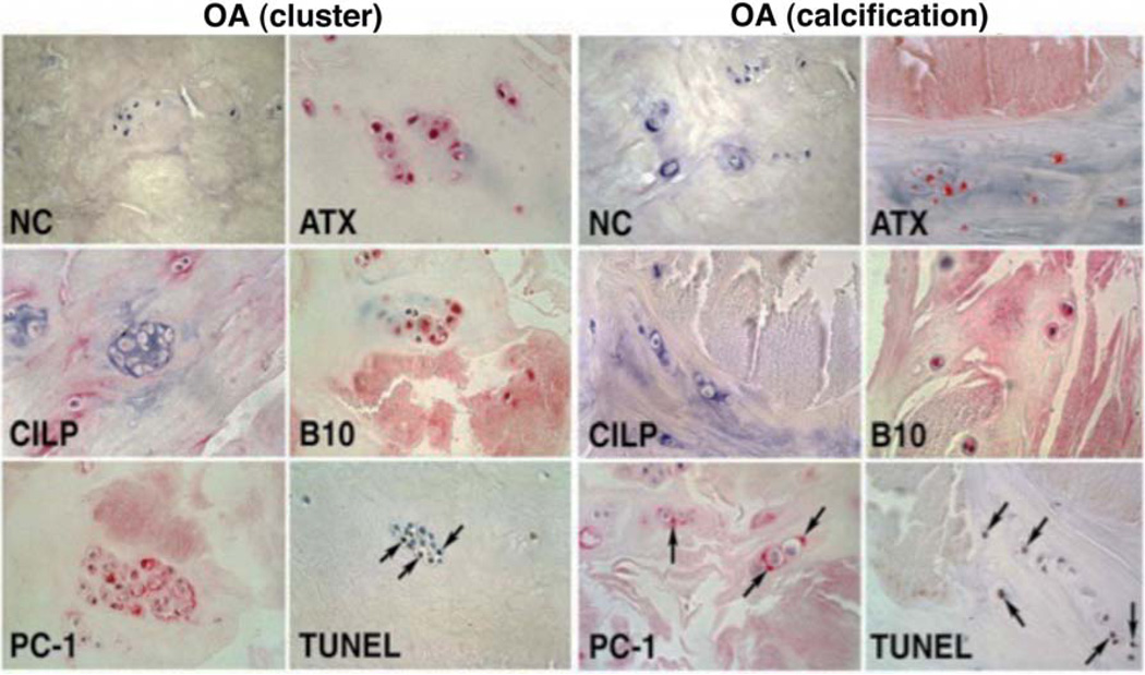
Figure 3. OA cluster cell apoptosis and calcification.
Localization of TUNEL-positive cells, calcium deposits and pyrophosphate-generating enzymes in menisci from OA-affected human knees. The left panel shows apoptotic cells, many in clusters, in the vicinity of (alizarin red-positive) calcified areas. The right panel shows cells immediately bordering calcifications. Staining for plasma cell membrane glycoprotein (PC-1), autotaxin (ATX) and B10 is also prominent at sites of calcification and in areas with TUNEL-positive cells. CILP: cartilage intermediate layer protein. Modified from (131).

