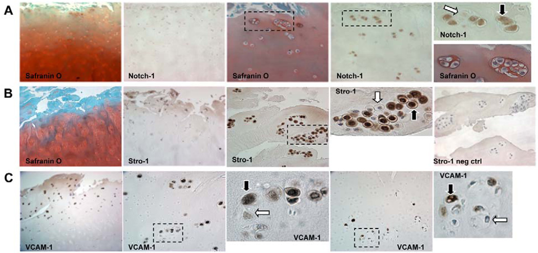
Figure 4. OA cluster staining for stem cell markers.
A majority of cells in clusters (69 to 79%) are positive for Notch-1, Stro-1 and VCAM-1. Clusters located in the DZ had significantly reduced frequencies of Stro-1 positive cells. (A) Safranin O and Notch-1 staining in clusters (×10 and ×40). (B) Safranin O and Stro-1 staining of OA cartilage sections (×10 and ×40). (C) OA cartilage sections immunostained for VCAM-1 (×10 and ×40). Positive staining indicated by black arrows and negative with white arrows. Modified from (138).

