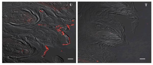
Figure 4. PNA-FISH magnified images (100x) of tongue tissue sections hybridized with a Cy3-conjugated C. albicans-specific probe.
Representative images demonstrating C. albicans presence on the tissue of untreated (U) tongues compared to presence on tongues treated (T) with Hst-5 or saliva. Bar represents 20μm.

