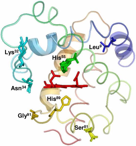
Figure 1.

Crystal Structure of DHP B (PDB accession code 3ixf). The location of the five residues (Leu9, Lys32, Asn34, Ser81 and Gly91) relative to the heme active site that differ in DHP A are shown, as well as the proximal (His89) and distal (His55) histidines.
