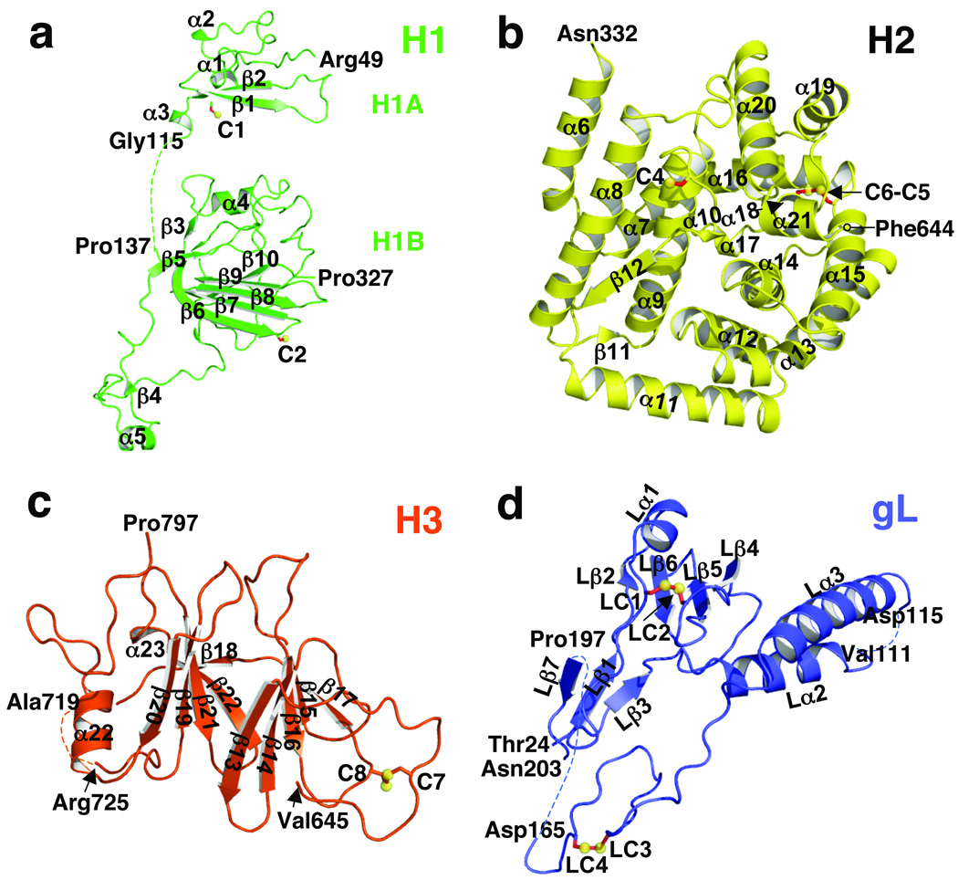
Fig. 2. Domains of gH and gL.
(a) domain H1, (b) domain H2, (c) domain H3, (d) gL. The coloring scheme is the same as in Fig. 1. Disulfides are shown as yellow spheres and red sticks and labeled. All secondary structure elements are labeled. Disordered segments are shown as dotted lines. Labeled residues indicate the limits of individual domains and the disordered loops.

