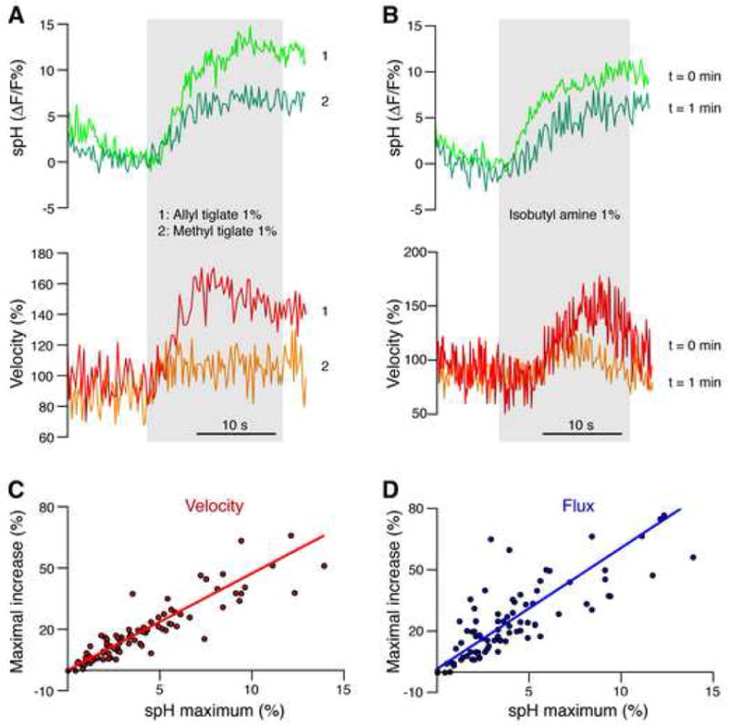
Figure 3. Presynaptic glutamate release and CBF are highly correlated.

(A) A glomerulus responded to two different odors with different amplitudes of spH fluorescence, as expected from different binding affinities of a given odorant receptor to different odors. The amplitude of CBF increase, shown as velocity changes during odor presentations, was related to presynaptic activity. The gray area indicates the duration of odor presentation.
(B) Measuring spH fluorescence in a glomerulus during repeated presentation of the same odor in short intervals revealed habituation of the presynaptic response, indicated by the smaller response to the second stimulus. A similar habituation pattern was observed in the velocity changes.
(C–D) Velocity and flux changes were highly correlated with presynaptic activity. Lines represent the Deming linear regression.
