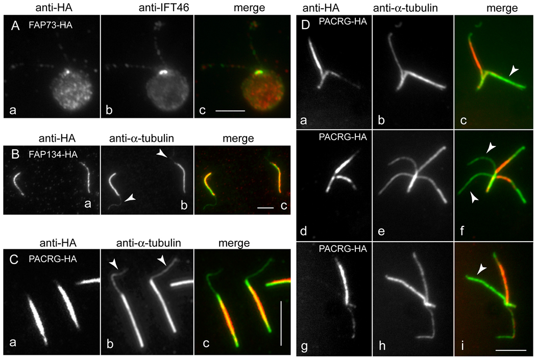
Fig. 2.
Co-localization studies of FAP73-HA, FAP134-HA, and PACRG-HA.
(A) Methanol-fixed cell expressing FAP73-HA stained with anti-HA (a) and anti-IFT46 (b). The merged image is shown in c. (B) Isolated flagella with protruding central pair (arrowheads in b) from a strain lacking endogenous FAP134 and expressing FAP134-HA, stained with anti-HA (a) and anti-tubulin (b). The merged image is shown in c. (C) Same as in B but from strain 4.3 expressing PACRG-HA. (D) Split axonemes of strain 4.3 expressing PACRG-HA, stained with anti-HA (a, d, g) and anti-tubulin (b, e, h). The merged images (c, f, i) show that staining representing PACRG-HA is strongly reduced or absent from some outer doublet microtubules. Strain 4.3 used for the images in C and D carries a PACRG RNAi construct in addition to the PACRG-HA vector. See Supplementary Fig. 1 for a more detailed characterization of this strain. Bars = 5 µm.

