Fig. 5.
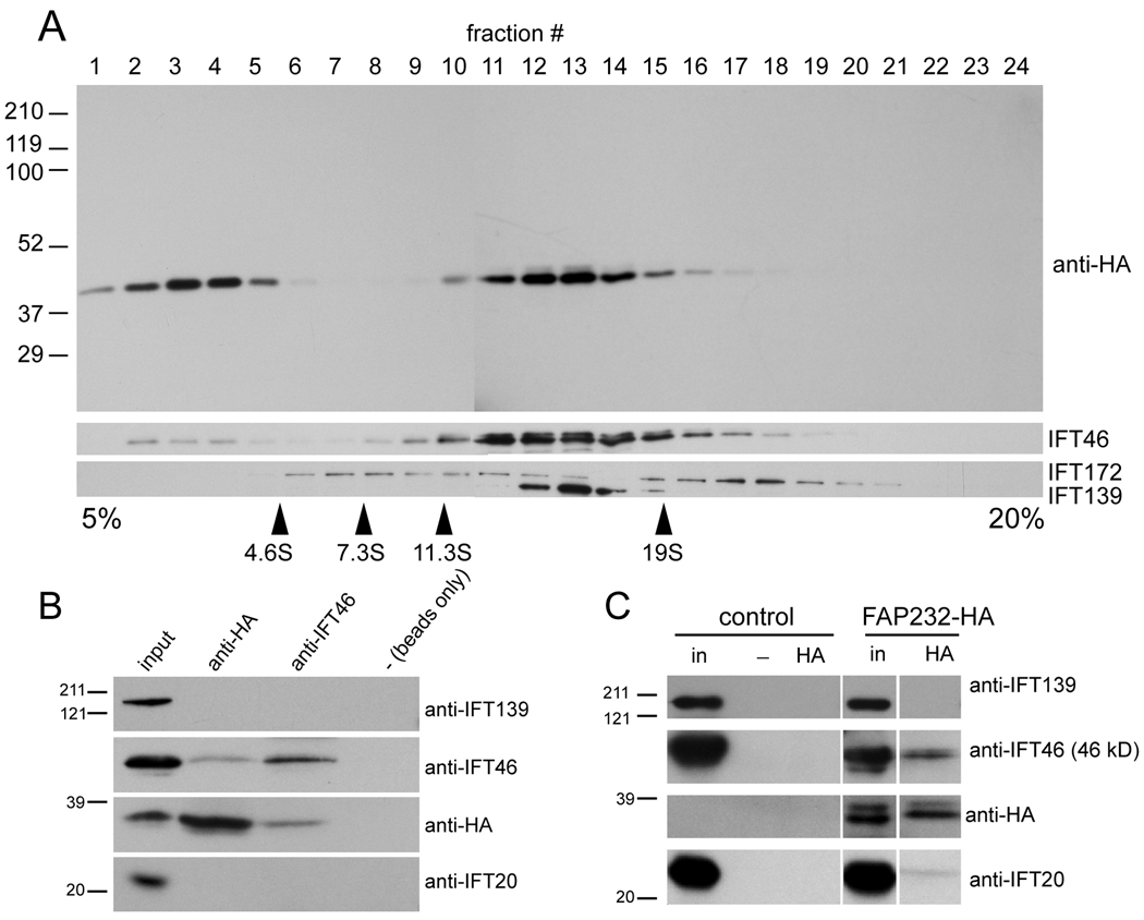
FAP232 is a component of IFT complex B. (A) Proteins in the membrane + matrix fraction obtained from flagella of cells expressing FAP232-HA were separated by sucrose density gradient centrifugation and analyzed by western blotting. The positions of marker proteins are indicated. FAP232-HA co-sediments precisely with complex B protein IFT46 but not complex A protein IFT139. (B) Western blot analysis of immunoprecipitates obtained from fractions 12 and 13 of the sucrose gradient shown in A. Protein G beads loaded with anti-HA antibody, protein A beads loaded with anti-IFT46 antibody, and protein G beads alone were used for the immunoprecipitations. The anti-HA antibody co-precipitated FAP232-HA and IFT46 but not IFT139. Similarly, the anti-IFT46 antibody co-precipitated IFT46 and FAP232-HA but not IFT139. The position of the marker proteins is indicated. (C) Western blots of immunoprecipitates obtained from membrane + matrix fractions from flagella of wild-type (control) and a strain expressing FAP232-HA, using anti-HA-loaded or unloaded (−) protein G beads. The anti-HA antibody co-precipitated FAP232-HA and complex B proteins IFT46 and IFT20, but not complex A protein IFT139.

