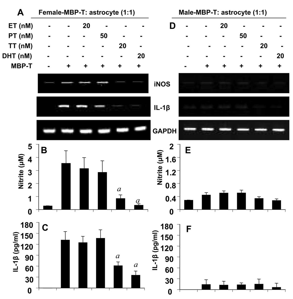
Figure 9. Effect of estrogen (ET), progesterone (PT), testosterone (TT), and dihydrotestosterone (DHT) on the ability of female and male MBP-primed T cells to induce contact-mediated expression of proinflammatory molecules in primary mouse astroglia.
Female (A – C) and male (D – F) MBP-primed T cells treated with respective concentrations of ET, PT, TT, and DHT for 72 h during MBP priming were added to astroglia at a ratio of 1:1 T cell:astroglia. After 1 h of stimulation, culture dishes were shaken and washed to lower T cell concentration. Then adherent astroglia were incubated in serum-free media for 5 h and the expression of iNOS mRNA was analyzed by semiquantitative RT-PCR (A & D). Adherent astroglia were incubated in serum-free media for 23 h and supernatants were used to assay nitrite (B & E) and IL-1β (C & F). Data are mean ± S.D. of three different experiments. a p < 0.001 vs. MBP-primed T cells only.

